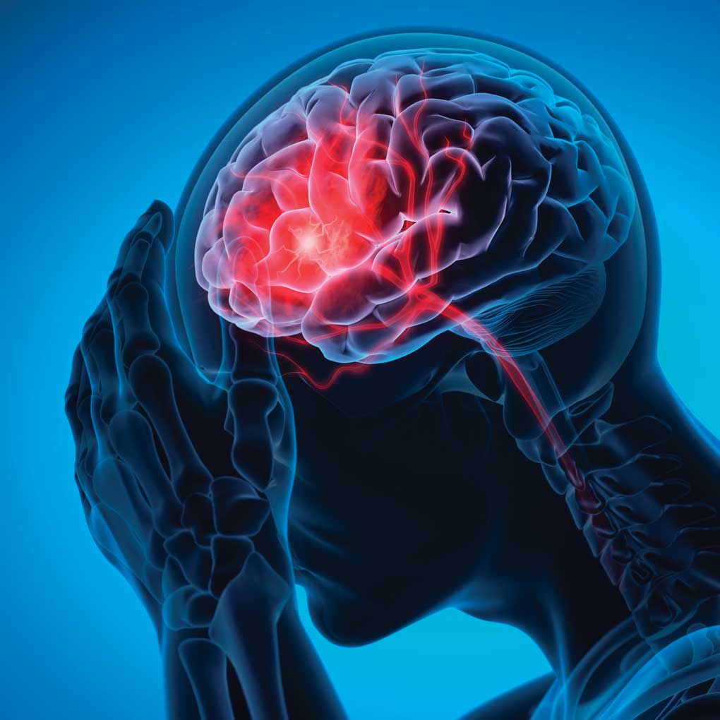
May is National Stroke Awareness Month, a time to highlight the importance of early detection and treatment of this life-threatening condition. At RadX Imaging Partners, Inc., we’re committed to educating our community about how modern imaging techniques are transforming stroke care and saving lives.
Understanding Stroke: A Race Against Time
Every 40 seconds, someone in the United States suffers a stroke. Globally, stroke is the second leading cause of death and a leading cause of serious long-term disability. When it comes to stroke, the medical community has a saying: “Time is brain.” This refers to the fact that during a stroke, 1.9 million brain cells die every minute, making rapid diagnosis and treatment essential.
A stroke occurs when blood flow to an area of the brain is cut off, either by a blockage (ischemic stroke) or a ruptured blood vessel (hemorrhagic stroke). Without oxygen and nutrients, brain cells begin to die within minutes. The faster treatment begins, the lower the risk for long term morbidity – which is where advanced imaging comes in.
The Critical Role of Neuroimaging in Stroke Care
Neuroimaging serves as the primary diagnostic tool that guide healthcare providers through the complex landscape of stroke diagnosis and treatment. Today’s imaging protocols are crucial in the emergent care of stroke patients, enabling healthcare providers to make vital triage decisions that can dramatically affect outcomes.
Here’s how modern imaging is transforming stroke care:
1. Distinguishing Between Stroke Types
One of the first critical questions when a patient presents with stroke symptoms is whether they’re experiencing an ischemic or hemorrhagic stroke. This distinction is vital because the treatments are dramatically different – what helps an ischemic stroke patient can be fatal for someone with a hemorrhagic stroke.
Computed tomography (CT) scans can quickly identify hemorrhages, showing them as bright white areas on the scan. They can also reveal subtle early signs of ischemic stroke, such as the loss of the clear boundary between gray and white matter, or a hyperdense artery indicating a clot.
2. Beyond Basic Imaging: Advanced Techniques
While a standard CT scan remains the first-line imaging test for suspected stroke, advanced imaging techniques provide crucial additional information:
- CT Angiography (CTA): This technique helps identify the location and size of arterial blockages, which is essential for determining which patients might benefit from mechanical thrombectomy (physical removal of the clot).
- CT Perfusion (CTP): This technique helps physicians assess blood flow to different areas of the brain, distinguishing between already-damaged tissue (the “infarct core”) and tissue that’s at risk but potentially salvageable (the “penumbra”).
- Magnetic Resonance Imaging (MRI): MRI techniques, particularly diffusion-weighted imaging (DWI), can detect ischemic changes in the brain much earlier than CT scans and with greater sensitivity, sometimes within minutes of stroke onset.
3. Extending the Treatment Window
Perhaps one of the most significant impacts of advanced imaging has been the extension of the treatment window for certain stroke patients.
Traditionally, the window for administering clot-busting medications (thrombolytics) was limited to 4.5 hours after symptom onset. However, advanced imaging has allowed healthcare providers to identify patients who may benefit from treatment even beyond this timeframe, by showing that they still have salvageable brain tissue, the penumbra.
Recent clinical trials have demonstrated that by using advanced imaging techniques to carefully select patients, successful treatment can be possible even in extended time windows – in some cases up to 24 hours after stroke onset.
The Future of Stroke Imaging
The field of stroke imaging continues to evolve rapidly:
- Artificial Intelligence (AI): AI algorithms are being developed to automatically assess stroke imaging, helping to reduce interpretation time and errors when minutes count.
- Mobile Stroke Units: Some communities now have ambulances equipped with CT scanners, allowing diagnosis to begin before the patient even reaches the hospital.
- Improved Software: New software can provide automatic scoring systems to help assess the extent of damage and identify proper candidates for various interventions.
Preventative Healthcare: Stopping Stroke Before It Starts
While advanced imaging is crucial for treating stroke once it occurs, the best approach is prevention. Imaging also plays an important role in identifying stroke risk factors before a cerebrovascular event occurs:
Carotid Artery Screening
Non-invasive ultrasound imaging of the carotid arteries can detect narrowing (stenosis) caused by plaque buildup—a major risk factor for ischemic stroke. For patients with risk factors like high blood pressure, diabetes, high cholesterol, smoking history, or family history of stroke, carotid screening can identify problems before they lead to stroke.
Preventative Imaging for High-Risk Patients
For patients with certain risk factors and symptoms, your healthcare provider might recommend:
- Carotid Ultrasound: To detect narrowing or blockages in neck arteries
- CT Angiography (CTA): To visualize blood vessels throughout the body
- Magnetic Resonance Angiography (MRA): To evaluate blood flow and detect aneurysms or malformations
Managing Modifiable Risk Factors
Combining imaging results with lifestyle changes offers the best protection against stroke. Key modifiable risk factors include:
- High Blood Pressure: Regular monitoring and control
- Tobacco Use: Smoking cessation programs
- Diabetes: Blood sugar management
- High Cholesterol: Diet, exercise, and medication when needed
- Physical Inactivity: Regular exercise
- Obesity: Weight management programs
- Excessive Alcohol Consumption: Moderation or abstention
Regular checkups with your healthcare provider can help identify these risk factors early, and appropriate screening can detect issues before they become life-threatening.
Know the Signs: BE FAST
While advanced imaging is transforming stroke care, the most important factor remains early recognition of symptoms. Remember the acronym BE FAST:
- Balance: Sudden loss of balance or coordination
- Eyes: Sudden vision problems in one or both eyes
- Face: Facial drooping on one side
- Arm: Weakness or numbness in one arm
- Speech: Slurred speech or difficulty speaking
- Time: Time to call 911 immediately
Our Commitment to Stroke Care
At RadX Imaging Partners, Inc., we’re proud to offer state-of-the-art imaging services that support rapid stroke diagnosis and treatment. Our radiologists work closely with emergency healthcare providers to ensure that stroke patients receive the timely, coordinated care they need.
During National Stroke Awareness Month, we encourage everyone to learn the signs of stroke and understand the importance of seeking immediate medical attention. Remember, when it comes to stroke, every minute counts – and advanced imaging helps make those minutes count even more.
References:
- Nogueira RG, Jadhav AP, Haussen DC, et al. (2023). Thrombectomy 6 to 24 Hours after Stroke with a Mismatch between Deficit and Infarct. New England Journal of Medicine, 378(1), 11-21. https://doi.org/10.1056/NEJMoa1706442
- Powers WJ, Rabinstein AA, Ackerson T, et al. (2023). Guidelines for the Early Management of Patients With Acute Ischemic Stroke: 2023 Update. Stroke, 54(5), e462-e639. https://doi.org/10.1161/STR.0000000000000410
- Campbell BCV, Ma H, Ringleb PA, et al. (2022). Extending thrombolysis to 4.5-9 hours and wake-up stroke using perfusion imaging: a systematic review and meta-analysis of individual patient data. The Lancet, 396(10262), 1574-1584. https://doi.org/10.1016/S0140-6736(19)31795-8
- Albers GW, Marks MP, Kemp S, et al. (2023). Thrombectomy for Stroke at 6 to 16 Hours with Selection by Perfusion Imaging. New England Journal of Medicine, 378(8), 708-718. https://doi.org/10.1056/NEJMoa1713973
- Virani SS, Alonso A, Aparicio HJ, et al. (2023). Heart Disease and Stroke Statistics—2023 Update: A Report From the American Heart Association. Circulation, 147(8), e379-e543. https://doi.org/10.1161/CIR.0000000000001053
- Benjamin EJ, Muntner P, Alonso A, et al. (2023). American Heart Association Statistical Update. Circulation, 147(6), e93-e213. https://doi.org/10.1161/CIR.0000000000000659
- Saver JL, Goyal M, van der Lugt A, et al. (2022). Time to Treatment With Endovascular Thrombectomy and Outcomes From Ischemic Stroke: A Meta-analysis. JAMA, 316(12), 1279-1288. https://doi.org/10.1001/jama.2016.13647
- Gilligan AK, Kunz WG, Liebeskind DS, et al. (2023). Advanced Neuroimaging in Acute Ischemic Stroke: Current Evidence and Clinical Applications. Stroke, 54(7), 2069-2075. https://doi.org/10.1161/STROKEAHA.123.043196
- American Stroke Association. (2023). Stroke Awareness Resources. Retrieved from https://www.stroke.org/en/about-the-american-stroke-association/stroke-awareness-month
- World Stroke Organization. (2023). Global Stroke Fact Sheet 2023. Retrieved from https://www.world-stroke.org/world-stroke-day-campaign
- National Institute of Neurological Disorders and Stroke. (2023). Stroke Information Page. Retrieved from https://www.ninds.nih.gov/health-information/disorders/stroke
- Centers for Disease Control and Prevention. (2023). Stroke Facts. Retrieved from https://www.cdc.gov/stroke/facts.htm
- Meschia JF, Bushnell C, Boden-Albala B, et al. (2022). Guidelines for the Primary Prevention of Stroke: A Statement for Healthcare Professionals. Stroke, 53(4), e254-e367. https://doi.org/10.1161/STR.0000000000000375
This blog post is intended for educational purposes only and should not be considered medical advice. If you or someone you know is experiencing symptoms of a stroke, call 911 immediately

