When it comes to breast health, early and accurate detection makes all the difference. That’s why medical professionals are increasingly turning to breast elastography—an advanced imaging technique providing crucial insights beyond traditional methods.
What Is Breast Elastography?
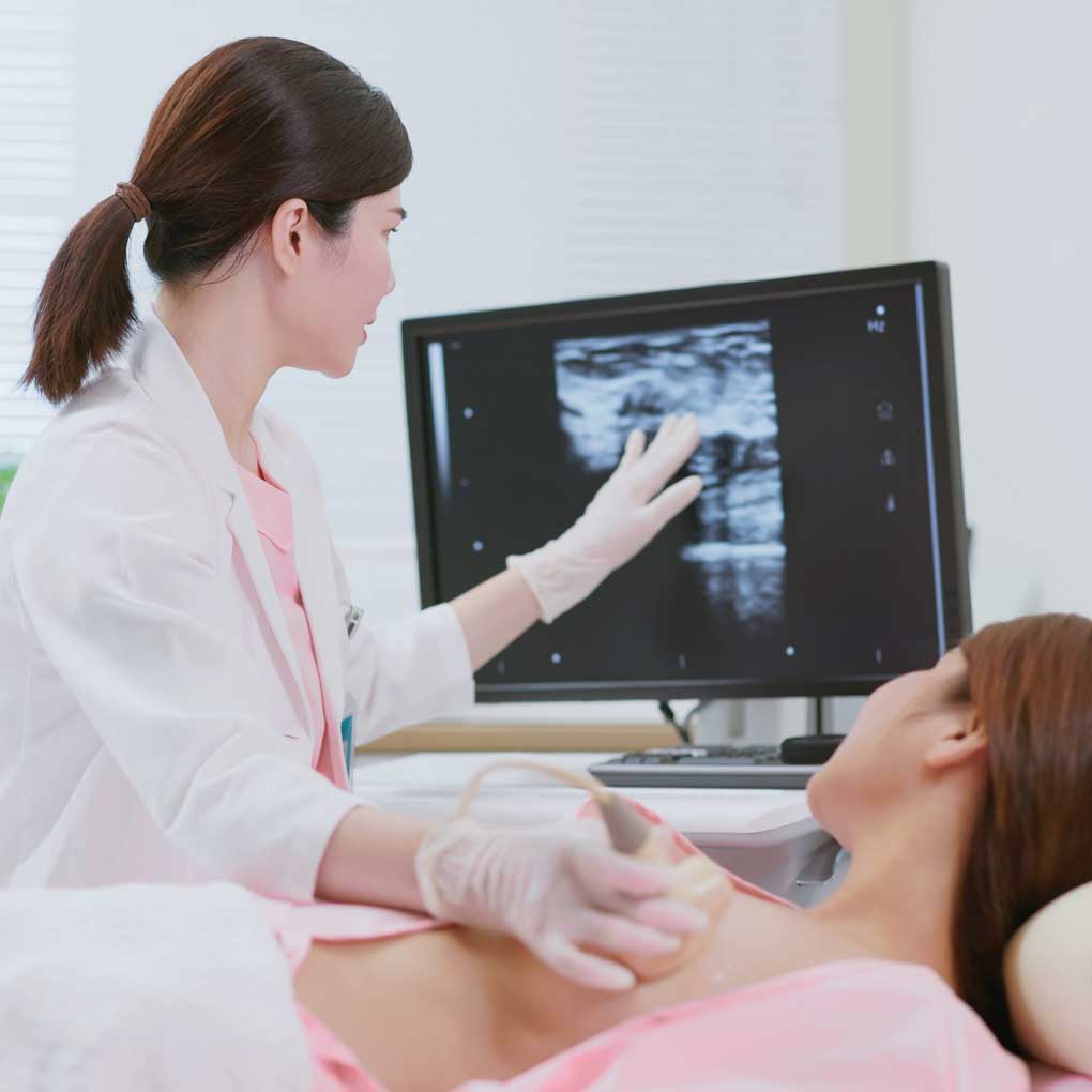
Breast elastography works like a specialized ultrasound that does more than just create images. It actually measures tissue stiffness—a critical factor since cancerous tissue is typically firmer than surrounding healthy tissue. This helps differentiate between soft, benign lesions and stiff, potentially malignant ones, particularly in cases where conventional ultrasound findings are inconclusive.
Medical providers use two main approaches:
- Strain elastography (SE): Measures how tissue deforms in response to external compression. Malignant tumors typically deform compared to benign lesions.
- Shear wave elastography (SWE): Uses acoustic radiation force to generate shear waves within tissues and measures their velocity. Higher velocity indicates increased stiffness, which is often associated with malignancy.
When Is It Recommended?
Your healthcare provider might suggest elastography when:
- A mammogram reveals an area of concern
- A traditional ultrasound shows an unclear result
- You have dense breast tissue that makes standard screening challenging
- Additional information is needed to determine if a biopsy is necessary
The Patient Experience
- Comfortable and quick: Similar to standard ultrasound
- No preparation required
- Non-invasive: No needles or radiation
- Immediate insights: Provides real-time information during your appointment
Clinical Applications
Differentiation of Benign and Malignant Lesions
Elastography significantly improves the differentiation of breast masses when used alongside conventional ultrasound. Malignant tumors exhibit higher stiffness values, whereas benign lesions remain relatively soft.
Reduction of Benign Biopsies
One of the key benefits is the potential to reduce benign biopsies. Conventional ultrasound alone, while a useful tool, is less effective at distinguishing between benign and malignant masses when compared to ultrasound in conjunction with elastography. Elastography adds an additional layer of confidence to the diagnostic process.
Complementary Role in Breast Cancer Screening
While mammography remains the gold standard for breast cancer screening, elastography serves as a valuable complementary tool, particularly for those with dense breast tissue where mammograms may be less effective.
Advantages and Limitations
Advantages
- Non-invasive and radiation-free
- Enhances diagnostic accuracy when combined with conventional ultrasound
- Reduces benign biopsies
- Can be used to monitor treatment response
Limitations
- Results can be operator-dependent, especially in strain elastography
- Variability based on different machines and settings
- May not be reliable for very deep-seated or very superficial lesions
The Science Behind It
Elastography has strong clinical evidence supporting its role in breast care. The BE1 multinational study of 939 masses demonstrated that shear-wave elastography can improve the specificity of breast ultrasound by up to 30% when added to conventional imaging, helping to reduce false positives and benign biopsies.
Next Steps
If you’re concerned about breast health or have been told you need additional imaging, ask about elastography at your next appointment. This technology represents an important advancement in how we approach breast health—bringing greater clarity, comfort, and confidence to your care.
Is Elastography Right for You?
If you’re wondering whether breast elastography might be beneficial in your specific situation, talk to your healthcare provider. They can evaluate your breast health history, risk factors, and previous imaging results to determine if elastography would provide valuable additional information in your case. Your provider can also discuss any limitations of the technology specific to your situation and help you make an informed decision about your breast health screening and diagnostic options.
Important Insurance Considerations
It’s important to note that breast elastography is not covered by all insurance payors or for all diagnoses. Coverage policies vary widely, and many insurers consider elastography a diagnostic rather than screening procedure. Before scheduling this imaging, check with your insurance provider about coverage specific to your plan and diagnosis. Some facilities offer cash-pay options for patients whose insurance doesn’t cover the procedure.
References
- Berg, W. A., Cosgrove, D. O., Doré, C. J., et al. (2012). Shear-wave elastography improves the specificity of breast ultrasound: The BE1 multinational study of 939 masses. Radiology, 262(2), 435–449.
- Chang, J. M., Won, J. K., Lee, K. B., Park, I. A., Yi, A., & Moon, W. K. (2013). Comparison of shear-wave and strain elastography in the differentiation of benign and malignant breast lesions. American Journal of Roentgenology, 201(3), W347-W356.
- Evans, A., Whelehan, P., Thomson, K., et al. (2018). Invasive breast cancer: Relationship between shear-wave elastographic findings and histologic prognostic factors. Radiology, 289(1), 45–52.
- Zhou, J., Zhan, W., Chang, C., et al. (2020). Breast lesions: Evaluation with shear wave elastography, with special emphasis on the “stiff rim” sign. Radiology, 274(1), 202–211.