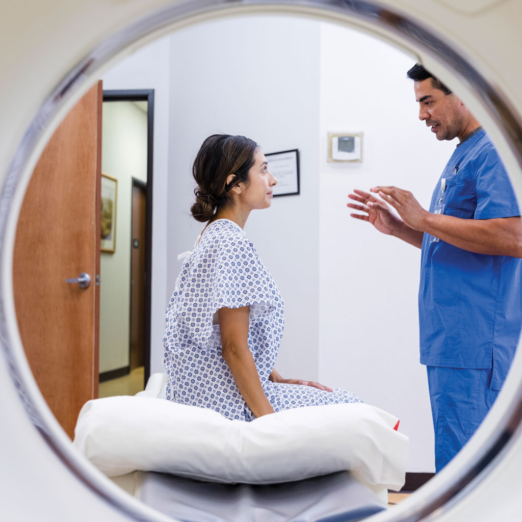
It is one of the most common questions patients ask, and it usually arrives sideways. How Much Radiation Is Actually in a Scan? Is this safe? Have I had too many of these? Should I be worried about my daughter having one?
The honest answer requires a unit of measurement most people have never used and a sense of scale that almost nobody carries around. This article supplies both.
The Unit
Radiation dose is measured in millisieverts, abbreviated mSv. The figure your radiologist would quote is called an effective dose, which accounts for the fact that different tissues respond differently to radiation, and it is designed so that exposures to different parts of the body can be compared on one scale.
It is an approximation rather than a measurement of what happened to you specifically. Actual dose varies with your size, with the equipment, and with how the exam was performed. The International Commission on Radiological Protection is explicit that effective dose has real limitations when applied to an individual patient, which is worth knowing before anyone treats a single number as precise.
The Scale You Already Live On
Everyone is exposed to radiation continuously, from the ground, from cosmic rays, and from radon in ordinary houses. According to the American College of Radiology and the Radiological Society of North America, the average person in the United States receives roughly 3 mSv per year from natural sources alone.
That number is not uniform. Living at altitude in Colorado or New Mexico adds about 1.5 mSv a year compared with living at sea level. Radon in the home is the single largest contributor, at roughly 2 mSv a year, and it varies enormously by region. A coast-to-coast round-trip flight is about 0.03 mSv.
None of that is something anybody worries about, and it is the right baseline for thinking about a scan.
Where Common Exams Fall
These are approximate adult figures published by ACR and RSNA, expressed both in millisieverts and as the equivalent amount of natural background radiation.
- Extremity x-ray, meaning a hand or a foot. Less than 0.001 mSv, under three hours of background.
- Bone density scan, DEXA. About 0.001 mSv, about three hours.
- Dental x-ray. About 0.005 mSv, about one day.
- Chest x-ray. About 0.1 mSv, about ten days.
- Screening digital mammogram. About 0.28 mSv, about 34 days.
- 3D screening mammogram. About 0.34 mSv, about 42 days.
- Lumbar spine x-ray. About 1.4 mSv, about six months.
- Low-dose CT for lung cancer screening. About 1.5 mSv, about six months.
- Cardiac CT for calcium scoring. About 1.7 mSv, about six months.
- CT of the brain. About 1.6 mSv, about seven months.
- CT of the chest. About 6.1 mSv, about two years.
- CT of the abdomen and pelvis. About 7.7 mSv, about 2.6 years.
The full published chart covers considerably more exams, and it is worth looking at the source directly rather than relying on a partial list.
Two patterns are worth pulling out of that. Plain x-rays sit far below a single year of background, often far below a single week. And CT is a different order of magnitude from x-ray, which is exactly why a physician thinks about whether a CT is the right test rather than ordering one reflexively.
What Has No Radiation At All
This gets lost in the anxiety. MRI uses a magnetic field and radio waves and involves no ionizing radiation. Ultrasound uses sound waves and involves none either.
That is a large part of why those two are preferred in pregnancy, in children, and in any situation where a patient will be imaged repeatedly over many years. If you are being followed for something long term, it is a reasonable question to ask your physician whether the follow-up can be done with one of them.
How Facilities Keep Dose Down
The governing principle is ALARA, meaning as low as reasonably achievable. In practice that means protocols matched to the specific clinical question rather than one setting for everyone, dose-reduction technology built into modern scanners, and pediatric protocols sized to the child rather than scaled down from an adult.
It also means not repeating a study that does not need repeating, which is one of the more underrated reasons to have prior imaging available for comparison. Bringing records from another facility, or authorizing their release in advance, sometimes eliminates a scan entirely.
The Question About How Much Radiation is Actually in a Scan That Matters
The risk from a single diagnostic exam is small. The risk from a missed or delayed diagnosis is frequently not small, and that comparison is the one that should drive the decision.
What is worth asking your physician is not whether the scan involves radiation, because you can look that up. It is whether this particular scan will change what happens next. If it will, the exposure is almost always justified. If it will not, that is a conversation worth having regardless of dose.
And if you are keeping track of your own imaging history, which is a reasonable thing to do, bring it up with your doctor rather than trying to total it yourself. Cumulative dose is a genuine consideration in some situations and a red herring in many others, and which one applies depends on your history.
References
American College of Radiology and Radiological Society of North America. Radiation Dose from X-Ray and CT Exams. Last reviewed April 15, 2025. radiologyinfo.org
International Commission on Radiological Protection. Publication 103. icrp.org
Image Gently Alliance. Radiation safety in pediatric imaging. imagegently.org