If your doctor has ordered MRI scans, you probably know it produces very detailed pictures of the inside of your body, and you may also know it involves a long tube and a lot of noise. Beyond that, most people are not sure what the exam actually involves or why the scheduler asked so many questions about metal. This article walks through it in plain terms.
What are MRI Scans?
MRI stands for magnetic resonance imaging. Unlike an X-ray or a CT scan, an MRI scans use no radiation at all. It works by combining a very strong magnetic field with radio waves, and a computer turns the resulting signals into detailed images that can be viewed from any angle.
What makes MRI scans different from other tests is how well it shows soft tissue. Muscles, ligaments, cartilage, the brain and spinal cord, the liver and other organs all show up with a level of detail that other imaging cannot match. That is why MRI is so often the test of choice for a knee injury, a back problem, or anything involving the brain.
What do MRI Scans Feel Like?
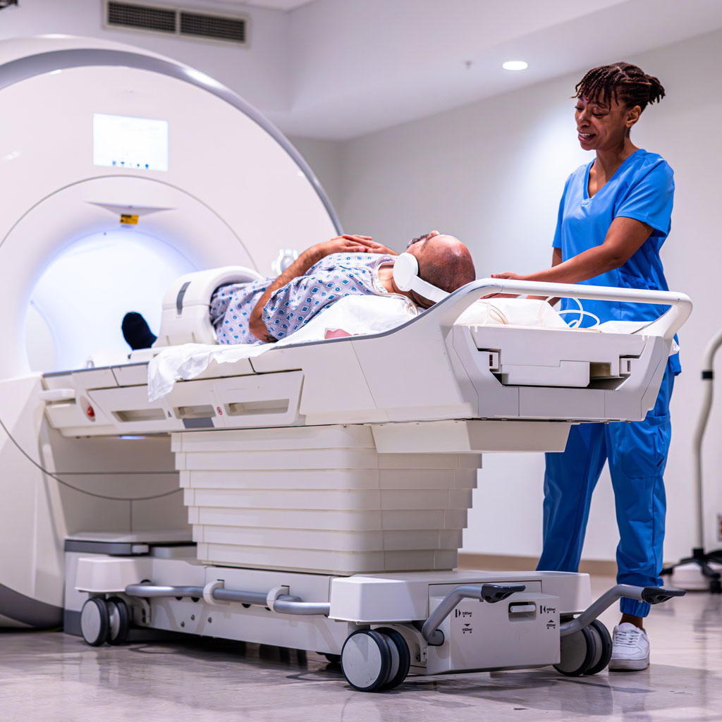
You lie on a padded table that slides into the scanner, and a technologist positions a device called a coil over or around the part of the body being imaged. The exam itself is painless, though most MRIs run longer than other scans, commonly 20 to 45 minutes depending on what is being studied and how many sequences your doctor ordered.
Two things surprise first-time patients. The first is the noise. An MRI is loud, with knocking and buzzing sounds that come and go throughout the exam, and you will be given earplugs or headphones. The second is how still you need to be. Motion blurs the images the same way it blurs a photograph, so short pauses between sequences are built in and the technologist talks to you through an intercom the whole way through.
Why the Safety Questions Matter
Before your exam you will be asked a long list of questions about metal in or on your body, and you will be asked again when you arrive. This is not paperwork for its own sake. The scanner’s magnet is always on, even between patients, and it is strong enough to move certain metal objects or to interfere with how an implanted device works.
Tell the scheduler and the technologist about any of the following, even if it was years ago and even if you think it is unrelated.
- A pacemaker, defibrillator, neurostimulator, cochlear implant, or any implanted pump
- Surgical clips, coils, stents, or a heart valve
- Joint replacements, plates, screws, or rods
- A metal fragment in the eye, or any history of work as a machinist, welder, or metalworker
- Body piercings, permanent makeup, or a medication patch
Many implants made today are safe in an MRI, and many are safe under specific conditions. The point of the questions is to identify the device and check it against the manufacturer’s information before you go anywhere near the magnet, which is also why bringing the implant card or the operative report helps so much.
Will I Need Contrast for my MRI Scans?
Some MRI scans use a contrast agent given through a small IV in the arm, which makes blood vessels, inflammation, and certain kinds of tissue stand out more clearly. Not every MRI needs it, and your doctor’s order specifies whether yours does.
If contrast is part of your exam, you may be asked about kidney function and about any previous reaction to contrast. Most people feel nothing beyond the IV itself, and a cool sensation in the arm during the injection is normal.
If Small Spaces Worry You
This is one of the most common concerns patients raise, and it is worth bringing up before the day of your exam rather than after you are on the table. Depending on what is being imaged, the opening may be shorter than you expect, your head may not need to go inside at all, and some scanners have a wider bore than others.
If you know that enclosed spaces are difficult for you or you experience claustrophobia, tell your referring physician when the exam is ordered. Many patients do well with a brief explanation of what will happen, a cloth over the eyes, or music through the headphones. Some do better with a mild medication prescribed by their own doctor, which needs to be arranged in advance and requires someone to drive you home.
How the Radiologist Reads MRI Scans
When the MRI scans are finished, the images go to a radiologist, a physician whose entire practice is interpreting medical imaging. An MRI study is not one picture but a set of sequences, each weighted to show different tissue properties, and the radiologist works through them in combination rather than one at a time. A finding that looks unremarkable on one sequence may be the key to the diagnosis on another.
The radiologist compares what the images show against your age, your history, and the clinical question your doctor asked, then writes a report that goes to your referring physician. If something needs attention right away, the radiologist calls your doctor rather than waiting for the report to be finalized.
What Happens After Your MRI Scans
There is no recovery period after routine MRI scans and you can eat, drive, and go back to work immediately. If you received contrast or took medication to help you relax, your care team will give you specific instructions before you leave.
Your doctor reviews the report and discusses the findings in the context of everything else they know about your health. If you have questions about what the report says, your referring physician is the right person to ask.
References
American College of Radiology. ACR Manual on MR Safety. acr.org
RadiologyInfo.org. Magnetic Resonance Imaging (MRI) Safety. radiologyinfo.org
U.S. Food and Drug Administration. MRI (Magnetic Resonance Imaging). fda.gov