Say the words ultrasound imaging and most people picture a pregnancy scan. That is a fair association, and it is a fraction of what the test actually does. Ultrasound is used on the gallbladder, the liver, the kidneys, the thyroid, the breast, the testicles, the heart, and the blood vessels, and it answers a wider range of questions than almost any other single test.
How Ultrasound Imaging Works
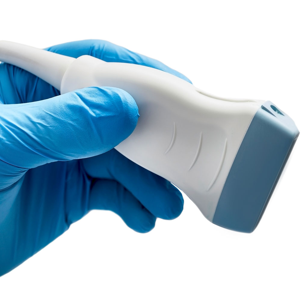
An ultrasound machine sends high-frequency sound waves into the body through a handheld probe and listens for the echoes that come back. Different tissues reflect sound differently, and a computer turns the timing and strength of those echoes into an image on the screen.
Two features follow from that. There is no radiation involved, which is why the exam is safe in pregnancy and can be repeated without accumulating any exposure. And the image is live, so the sonographer watches structures move in real time rather than capturing a single frozen moment.
What Ultrasound Imaging Is Like
A sonographer applies a warm gel to the skin, which eliminates the thin layer of air that would otherwise block the sound waves, and moves the probe over the area being studied with a moderate amount of pressure. Most exams take 20 to 45 minutes.
You will be asked to hold your breath at points, and possibly to turn on your side or change position, because the angle of the probe determines what can be seen. The exam is not painful, though pressing over an area that already hurts can be briefly uncomfortable, and that reaction is itself diagnostic information.
Preparation depends on what is being imaged. A gallbladder or abdominal study usually requires fasting, because a gallbladder that has recently emptied is difficult to evaluate. A pelvic study often requires a full bladder. A thyroid, breast, or vascular study usually requires nothing at all. Follow the instructions you were given, since a poorly prepared exam frequently has to be repeated.
Where Is Ultrasound Imaging the Right Answer
Ultrasound is not simply the cheaper option. For a number of questions it is the best test available.
For gallstones and gallbladder inflammation it outperforms CT. For distinguishing a simple cyst from a solid nodule in the thyroid or the breast it is excellent and it is fast. For blood clots in the legs it is the standard first test, using Doppler to show the direction and speed of blood flow and whether a vein compresses normally. In breast imaging it is what characterizes something seen on a mammogram, and it is a standard supplemental tool for women with dense tissue.
Where Ultrasound Imaging Has Limits
Sound does not travel through air or bone, which means ultrasound cannot see the lungs well, cannot see the brain through an adult skull, and has trouble looking past bowel gas. Depth is a constraint as well, so structures far below the surface are harder to resolve.
Ultrasound is also more operator-dependent than other imaging. The images are acquired by a person making judgments in real time about angle, pressure, and what deserves a closer look, which is why the training and experience of the sonographer and the radiologist reading the study matter more here than in almost any other modality.
Who Reads Ultrasound Imaging
The sonographer performs the exam and captures the images. A radiologist then interprets them, integrates them with your history and the clinical question, and writes the report that goes to your referring physician. If a finding requires prompt attention, the radiologist contacts your doctor directly rather than waiting for the report to be finalized.
Afterward
There is nothing to recover from. You wipe off the gel, get dressed, and go about your day, and if you fasted for the exam you can eat immediately. Your doctor reviews the report and discusses what it means alongside everything else they know about your health.
References
RadiologyInfo.org. General Ultrasound. radiologyinfo.org
American Institute of Ultrasound in Medicine. Practice Parameters. aium.org
American College of Radiology. ACR Appropriateness Criteria. acr.org